Prostate CT is an instrumental examination aimed at identifying or clarifying pathology. This is a fairly universal diagnostic method with many advantages, although it also has some contraindications. As a result of its implementation, a conclusion is drawn up, with the help of which a specialized doctor is able to diagnose the pathology.
What does a CT scan show?
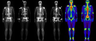
Computed tomography of the prostate is one of the modern methods of examining this organ for the purpose of diagnosing neoplasms and other pathologies. Using X-rays, a three-dimensional image of the organ and vascular-lymphatic system is created. This allows you to evaluate the structural features, density, contours, angle, condition of the seminal vesicles, uniform distribution of fatty tissue and the ratio of the parameters of the prostate gland to the bladder and other pelvic organs.
CT makes it possible to identify the following pathologies:
- Traumatic organ damage.
- Foreign bodies.
- Allows you to show prostate cancer.
- Adenoma.
- Inflammatory processes.
- Lymphadenopathy during the oncological process.
- Circulatory disorders and bleeding.
The advantages of diagnosing using computed tomography are:
- Painless and non-invasive (does not require surgical or other procedures).
- High speed and ease of examination.
- Availability.
- Highly informative (the information obtained can be used by doctors of various profiles when diagnosing pathologies).
Indications
Computed tomography is indicated for:
- decreased quality of sexual life;
- disturbance and pain when urinating;
- suspected infertility;
- diagnosing tumors, including cancer and their possible spread to other organs of the genitourinary system in men;
- planning radiation therapy;
- detection of regional lymphadenopathy.
Contraindications
Carrying out a CT scan of the prostate gland cannot cause harm to health, so there are practically no contraindications. There may be some restrictions due to the different design of the devices themselves, for example, weight restrictions, because most tomographs cannot accommodate people with a large body weight (about 150 kg). Also, people with poor coordination may be given sedatives before the procedure.
Given that the device uses X-rays, there is some risk of radiation exposure. But this only applies to those being examined repeatedly, because a single dose of radiation received during an examination is insignificant. Therefore, it is recommended to take a break of 3-4 weeks between repeated procedures.
There are some contraindications for CT with contrast - it is not prescribed for people with allergies or intolerance to contrast, for people with heart or kidney failure, for diabetes or thyrotoxicosis.
Preparing the patient for CT
Conducting a computed tomography scan with contrast requires preparatory measures 2 days before the examination. You should eat only dietary foods and exclude from your diet foods that can increase gas formation (they may additionally prescribe medications that prevent bloating, for example, Espumisan). The day before the procedure, you must take laxatives and give an enema to cleanse the intestines. You should also refrain from eating on the day of the examination (last meal at least 6 hours before the procedure). You should come for diagnostics in comfortable clothes (because the procedure takes from 10 to 30 minutes) without metal elements.
In case of exhaustion or dehydration, which is possible with chronic diseases or prostate cancer, intravenous drips with saline and plasma-substituting solutions are prescribed to reduce the negative effects of contrast and accelerate its removal from the body. This procedure is carried out another day after the examination.
Antihistamines may also be prescribed a few days before the procedure, and corticosteroids may be administered on the day of the procedure.
How does a CT scan work?
The patient is placed on the table and placed in the tomograph, after which the examination begins. At the same time, all strangers leave the room. The main element of the tomograph in the form of a ring rotates around the patient’s area being examined, it is quite noisy, sometimes a crackling sound is possible, this should not be alarmed.
Contrasting to Increase Efficiency
Often, CT scans are performed using contrast enhancement; this increases the accuracy of observation and allows for a better study of the structure of the organ being examined and its circulatory system. Contrast is a non-toxic substance with a high iodine content, which is independently eliminated from the body within 1-2 days.
Contrast is injected intravenously before a CT scan and stains the blood vessels within a minute.
An experienced doctor will be able to give a preliminary assessment or identify the problem within the first minutes of the examination.
Interpretation of CT results
After the scan, the radiologist interprets the results and draws up a conclusion that will be necessary for specialized doctors to diagnose pathologies and prescribe treatment.