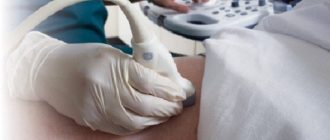
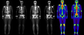
Prostate MRI is an instrumental method for diagnosing prostate pathologies. There are several ways to perform magnetic resonance imaging. All of them are preceded by a short preparatory period, during which it is necessary to monitor nutrition and adhere to simple rules. The diagnosis itself takes from 30 to 90 minutes, after which the specialist draws up a conclusion on the basis of which treatment is prescribed.
What does an MRI show?
MRI of the prostate in men shows the presence of prostatitis, abscess, adenoma and oncological processes. The scan allows you to determine the structure, size and position of the gland, as well as determine the presence and type of pathology.
In the diagnosis of cancer, adenoma and prostatitis, MRI of the prostate has the highest accuracy compared to other examination methods, especially when combined with a biopsy. A large-scale study was conducted at the Australian Wesley Institute to compare the effectiveness of existing diagnostic methods. The experiment involved almost 500 patients with elevated PSA levels. Each of them was examined by all four methods: ultrasound, TRUS, MRI with and without biopsy. Almost half were diagnosed with prostate cancer. Ultrasound showed the lowest accuracy - 62%, TRUS was slightly better - 77%. MRI without biopsy showed an accuracy of 89%, with biopsy - 100%.
The fact is that when conducting magnetic resonance imaging in parallel with a biopsy, it is possible to take material not blindly, but specifically from those areas that look like tumors. This allows you to reduce possible complications and the number of punctures from 30-40 to 10-12.
Indications
MRI of the prostate is done after undergoing simpler examinations, if as a result of them it was not possible to diagnose the presence or absence of pathologies (this is due to the inaccessibility of this type of examination). Indications for MRI of the prostate are:
- increased PSA in the blood, which may indicate oncological diseases - cancer or adenoma (visible as nodes in the gland);
- venereal diseases;
- infectious or fungal diseases;
- inflammatory diseases – acute and chronic prostatitis;
- stones in the prostate;
- complications after surgical interventions;
- examination before surgery.
In addition to examining the organ itself, the peripheral zone of the prostate, bladder, testicles, as well as nerve and bone tissue in the pelvis are examined.
Contraindications
MRI of the prostate is contraindicated in the following cases:
- Allergy to contrast agents (gadolinium, reaction testing occurs before the examination).
- Having a pacemaker, insulin pumps or metal implants.
- Surgical clips.
- Hearing aid.
- Claustrophobia.
- Large body weight (if present, diagnostics can be carried out on an open device without side walls).
Preparing for an MRI
Preparation for the study requires the implementation of certain restrictions and procedures:
- Refusal of foods that contribute to gas formation in the intestines (cabbage, carrots, legumes, cereals, carbonated and fermented milk products).
- Refusal to eat and 6 hours before the examination.
- Performing an enema before performing it.
- Removing jewelry and other metal objects from the body.
- Empty your bladder before the examination; you should not drink water after this.
- If there is increased anxiety, the patient may be prescribed mild sedatives.
How is an MRI of the prostate performed and how long does it take?
The examination may differ depending on the tomography method:
- Traditional method.
- With contrast.
- Multiparametric tomography.
Traditional MRI method
It is carried out relatively rarely, because the accuracy is inferior to the method using a contrast agent. But, if the patient has an allergic reaction to the contrast, the traditional method is used. The patient is immersed in the tomograph up to his waist. The procedure takes from 30 to 60 minutes. During this time, about 20 pictures are taken with a break of 2-3 minutes. During the scan you need to lie down without moving, during breaks you can move a little, the radiologist will additionally warn you about this.
With contrast
MRI of the prostate with contrast provides more accurate results than traditional ones. Before immersion in the tomograph, the patient is injected intravenously with a contrast agent based on gadolinium salts. Within a few minutes, the drug spreads through the blood and reaches the prostate, which allows you to obtain a more accurate image when scanning with a tomograph. The drug leaves the body naturally within 1-2 days.
Multiparametric MRI
Multiparametric MRI of the prostate with contrast is performed on a more highly accurate machine than with previous methods. The procedure itself also lasts 20-30 minutes longer, and deciphering the results requires more time. The advantages of this method are even higher accuracy, but without the use of a biopsy.
Interpretation of prostate MRI results
Performed by the same specialist who performed the scan. During decoding, the doctor identifies areas with pathologies and draws up a conclusion. It must be referred to the attending urologist, who, based on it, will make a diagnosis and prescribe treatment.


