Prostate biopsy is a universal diagnostic procedure that can accurately determine the course of the oncological process in the prostate. There are several techniques and methods of implementation, they differ in accuracy and method of implementation. Before the procedure, a preparatory period with simple restrictions is indicated, which reduces the likelihood of complications after it. The result is produced after 7-10 days.
What is it like?
A puncture biopsy of the prostate gland is a study whose purpose is to extract parts of the functional tissue of the organ with a needle. Next, a cytological and histological study of their structure and possible pathologies is carried out. Currently, it is a universal method for diagnosing benign and malignant prostate tumors in men.
Types and methods of carrying out
Puncture of the prostate gland can be performed in several ways, which differ in the technique of the procedure and the puncture sites.
Prostate biopsy is classified according to the sampling route into:
- Transrectal - the needle is inserted through the rectum.
- Transurethral – performed through the urinary tract.
- Transperineal - through an incision in the perineum.
The following biopsy methods are possible:
- Sextant puncture of the prostate - material is taken from 3 areas of the prostate, on both sides (a total of 6 biopsies are taken). Usually performed transrectally, but in case of rectal complications it can be performed transperineally. Diagnostic accuracy is about 40%.
- Multifocal biopsy (also called polyfocal or extended) - has no differences from the sextate method, in addition to the fact that the number of samples taken increases to 10-18, which makes it possible to more accurately study the condition of the organ. Performed under transrectal ultrasound guidance. Diagnostic accuracy is about 60%.
- Saturation biopsy - the number of samples taken depends on the PSA level and the size of the gland from 8 to 24. It is performed under TRUS control and only transperineally. Diagnostic accuracy is 70-80%.
- Fusion biopsy (also called targeted biopsy) is the most modern technique that involves the use of a three-dimensional model of the prostate created as a result of an MRI examination. It is superimposed on the ultrasound image, which allows you to take samples precisely. As a standard, 18 biopsies are taken and performed transperineally. Diagnostic accuracy is 80-90%.
Previously, puncture was performed with a thin needle and was called aspiration. But, with the development of medical technologies, the procedure began to be performed with a special high-speed needle and became known as trephine biopsy. The aspiration method is now practically not used due to increased pain and frequency of complications.
Indications
A prostate biopsy is done in cases of suspected benign (adenoma) or malignant (cancer) tumor. This procedure is usually prescribed after simpler tests and serves as a definitive way to make a diagnosis.
Indications for prostate puncture are as follows:
- increased total PSA level (above 4 ng/ml) or decreased ratio of free PSA to total (below 15%);
- detection of compacted areas during palpation of the gland;
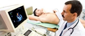
- detection of areas with low echogenicity using TRUS;
- control of the disease after TUR (transurethral resection of the prostate) or removal of the gland.
A repeat biopsy may be ordered:
- to clarify the stage of the disease for already diagnosed cancer;
- PSA levels remain elevated after therapy;
- During the initial puncture, prostatic intraepithelial neoplasia was detected;
- During the initial collection of material, it turned out to be insufficient for research.
Contraindications
The procedure is not prescribed in the following cases:
- puncture cannot be done on an inflamed prostate (in case of acute prostatitis);
- general serious condition of the patient;
- violation of the blood clotting mechanism;
- acute infectious diseases;
- for acute inflammatory diseases of the rectum or hemorrhoids.
A biopsy should not be done too often, as this can cause complications; the need for its appointment is considered by the doctor individually. In some cases, the procedure has to be abandoned due to the patient’s categorical disagreement with this type of diagnosis.
Preparation
In many ways, the procedure is similar to minimally invasive surgery, so you need to prepare for prostate puncture. When prescribing a diagnosis, the doctor will conduct a consultation, during which he will talk in detail about all restrictive measures and prescribed medications, and may give some individual advice, but in general, the preparatory procedures are as follows:
- Consultation and examination with a doctor for sensitivity to anesthetics and other factors that can cause complications.
- Before a prostate biopsy, you should not take anticoagulants (blood thinners) for at least a week. For 3 days – you cannot take non-steroidal anti-inflammatory and hormonal drugs. If it is impossible to completely refuse them, the doctor will prescribe analogues.
- For a week you are required to adhere to a dietary diet (the diet should not contain fatty foods, foods that complicate digestion, coffee and alcohol). If the examination takes place under anesthesia, you will need to completely abstain from food for 8-12 hours.
- Mild sedatives may be prescribed to reduce anxiety a few days before the diagnosis.
- Antibiotics are prescribed and continued after the examination.
- For transrectal or transurethral puncture, a cleansing enema is given before the procedure.
How to do a prostate biopsy
Prostate biopsy is performed in different ways, depending on the method chosen. A common feature is the use of antibiotics before the procedure. They are necessary to prevent possible contamination during sample collection.
Transrectal method
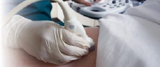
Transrectal puncture in men takes about 20-25 minutes. To do this, the subject is seated in a chair and fixed in a position convenient for taking samples. The procedure takes place under TRUS control, which allows you to take material with greater accuracy (especially if you have an MRI of the prostate). In some cases, anesthesia may be used, this is decided individually at the discretion of the surgeon. Samples are taken using a flexible probe. 10-12 samples are taken.
Prostate biopsy through the rectum is the least harmful and painful, therefore it is used most often.
Transurethal method
Transurethral puncture of the prostate is performed over 30 minutes. With this method, a cystoscope is inserted into the urethral canal, at the end of which there is a video camera and a cutting loop, which is used to take scrapings of organ tissue. Anesthesia is used at the discretion of the doctor.
Transperineal collection
The most rare way to take a prostate biopsy in men. First of all, local anesthesia of the area is performed using gel or injections or general anesthesia, after which an ultrasound machine is inserted transrectally. Next, about 20 samples are taken using a special device with needles. The whole process lasts 30-40 minutes.
Possible consequences
If you follow all the rules and recommendations of doctors, the harm from a biopsy is minimal, the risk of developing consequences and complications is minimal. The following manifestations are possible:
- discomfort in the rectum;
- elevated temperature;
- difficulty urinating;
- presence of blood in urine, feces and semen;
- the most dangerous phenomenon is infection and inflammation.
Almost all consequences go away on their own without intervention within 3 days; if this does not happen, you must consult your doctor.
To minimize the negative consequences, a regimen is prescribed that includes restrictions on physical activity, sex and diet.
results
The results of the prostate biopsy analysis are made 7-10 days from the moment the samples are taken. Decoding occurs according to the Gleason table, the condition of the organ is assessed on a 5-point scale:
- The neoplasm is a single cluster, the cell nuclei are not changed.
- The neoplasm is a small cluster that is separated by a membrane from healthy tissue.
- The neoplasm consists of a large accumulation of cells that grows into healthy tissue.
- The neoplasm consists of modified cells.
- The neoplasm consists of many modified cells that have grown into healthy tissue.
In addition to the table, the results are evaluated according to the Gleason index - the sum of the 2 highest results from all samples, which increases the diagnostic accuracy. The Gleason index is deciphered as follows:
- 2-4 – slow-growing oncological process;
- 5-7 – moderately aggressive oncology;
- 8-10 – fast-growing cancer with a high probability of metastasis.